Blog post
Dec 4, 2025

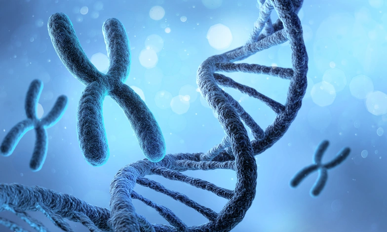
Chromosomes contain much of our genetic information and play an essential role in the development and functioning of the human body. When there is a change in their number or structure, this is known as a chromosomal abnormality, which can have very different consequences depending on the chromosomes involved.
These abnormalities may be associated with certain genetic conditions, fertility problems or pregnancy loss. However, not all chromosomal changes cause symptoms or affect health in the same way.
Understanding the different types of chromosomal abnormalities and the tests available to identify them can help people interpret genetic results and make informed decisions about their health and reproductive options.
Chromosomal abnormalities are changes in the number or structure of chromosomes. They can be numerical, such as trisomies and monosomies, or structural, such as translocations and deletions. They can be identified through genetic tests, including karyotyping, prenatal diagnostic testing and, in certain fertility treatments, preimplantation genetic testing of embryos.
In this article you will learn:
A chromosomal abnormality is a change affecting the number or organisation of chromosomes within a cell.
Most human cells normally contain 46 chromosomes arranged in 23 pairs. Of these, 22 pairs are autosomes, while the remaining pair consists of the sex chromosomes, usually XX or XY.
Each chromosome contains DNA organised into genes that provide essential instructions for the body's development and functioning.
When extra chromosomal material is present, genetic material is missing or chromosomes are rearranged, these changes can affect development or certain biological functions.
Chromosomal abnormalities may arise during the formation of eggs or sperm, develop during the earliest stages of embryo development or, in some cases, be inherited from a parent.
Not all chromosomal abnormalities necessarily cause a medical condition. Some chromosomal rearrangements do not produce symptoms in the people who carry them, although they may have reproductive implications. Others can cause significant developmental abnormalities.
Their clinical significance therefore depends on the type of change, the chromosomes involved and the individual's medical circumstances.
Chromosomal abnormalities are generally divided into two main categories: numerical and structural. Understanding this distinction helps explain what has changed in the genetic material and the potential consequences.
Numerical chromosomal abnormalities occur when a cell contains a different number of chromosomes from the usual number.
One of the most common forms is aneuploidy, which involves the presence or absence of one or more individual chromosomes.
The main types include:
Aneuploidies can arise when chromosomes fail to separate correctly during cell division, a process known as nondisjunction.
Some aneuploidies are compatible with live birth, while others interfere with embryo development and may result in miscarriage.
Changes involving entire additional sets of chromosomes, such as triploidy, can also occur. These are distinct from aneuploidies affecting individual chromosomes.
Structural chromosomal abnormalities occur when the organisation of one or more chromosomes changes, even though the total number of chromosomes may remain the same.
The main types include:
An important distinction is whether these rearrangements are balanced or unbalanced.
In a balanced rearrangement, genetic material is reorganised without a significant overall gain or loss. A person may carry such a rearrangement without experiencing symptoms and only discover it during fertility investigations.
By contrast, unbalanced rearrangements involve the loss or duplication of genetic material and may have more significant clinical consequences.
For example, someone carrying a balanced translocation may be healthy but have an increased risk of producing embryos with an unbalanced chromosomal complement.
Some chromosomal abnormalities are associated with well-known syndromes that have specific clinical features and healthcare needs. However, the effects can vary considerably between individuals, even when they share the same chromosomal diagnosis.
Down syndrome is a genetic condition usually associated with the presence of an additional copy of chromosome 21.
Most cases are caused by complete trisomy 21, although some involve chromosomal translocations or mosaicism.
It may be associated with certain physical characteristics, varying degrees of intellectual disability and an increased likelihood of medical conditions such as congenital heart defects or thyroid disorders.
However, the abilities, experiences and healthcare needs of people with Down syndrome vary considerably.
Most cases occur spontaneously during the formation of eggs or sperm. The likelihood of trisomy 21 increases with maternal age, although it can occur in pregnancies at any age.
Prenatal screening tests can estimate the likelihood of Down syndrome, while diagnostic tests can confirm the chromosomal finding.
Edwards syndrome and Patau syndrome are chromosomal conditions caused by an additional copy of chromosomes 18 and 13, respectively.
This condition is commonly associated with significant growth and developmental abnormalities, as well as congenital malformations that may affect the heart and other organs.
Many affected pregnancies end in miscarriage or stillbirth. Babies born alive often face serious medical complications, although individual outcomes can vary.
Patau syndrome can also cause significant abnormalities during fetal development, including malformations affecting the brain, heart and other organs.
As with trisomy 18, there is a high risk of pregnancy loss and serious complications after birth.
Both trisomies may be identified through prenatal screening and confirmed by diagnostic testing.
Support from specialists in genetics and fetal medicine is particularly important to help families understand the findings and the available care options.
Turner syndrome is a chromosomal condition affecting females, associated with the complete or partial absence of one X chromosome.
The classic form is known as monosomy X, represented by the karyotype 45,X. However, other forms exist, including mosaicism, in which different cells have different chromosomal compositions.
Common features include short stature, differences in ovarian development and absent or irregular menstrual periods.
Some individuals may also have cardiovascular, kidney or endocrine conditions requiring specialised medical monitoring.
Reproductive potential varies from person to person. Many women with Turner syndrome experience ovarian insufficiency, although some, particularly those with certain mosaic forms, may retain ovarian function.
Importantly, pregnancy in women with Turner syndrome requires a specialist cardiovascular assessment before conception, as pregnancy can carry significant health risks.
Klinefelter syndrome is a sex chromosome condition affecting males. In its classic form, an additional X chromosome is present, resulting in a 47,XXY karyotype.
Symptoms may be mild or go unnoticed during childhood. In some cases, the diagnosis is not made until adolescence or adulthood.
Possible features include smaller testicular volume, reduced testosterone production, differences in pubertal development and fertility difficulties.
For some men, infertility is the first sign that leads to a diagnosis.
However, Klinefelter syndrome affects individuals differently. Hormone production and the possibility of retrieving sperm can vary according to individual circumstances.
Karyotyping can identify this condition and help guide any additional medical investigations or reproductive counselling that may be appropriate.
Maternal age is one of the most important factors associated with the occurrence of certain numerical chromosomal abnormalities in embryos.
Egg cells begin developing before birth and remain in the ovaries for many years. Over time, the mechanisms responsible for ensuring that chromosomes separate correctly during egg maturation may become less reliable.
This increases the likelihood of chromosome segregation errors, which may result in eggs containing additional or missing chromosomes.
If one of these eggs is fertilised, the resulting embryo may have an aneuploidy.
This helps explain why the likelihood of certain trisomies and pregnancy losses associated with chromosomal abnormalities increases with maternal age.
However, several points are important to understand:
Maternal age is therefore a risk factor, not a diagnosis. Personal medical history and reproductive circumstances are essential when deciding whether additional genetic investigations may be appropriate.
Different tests can be used to examine chromosomes, but they do not all serve the same purpose. Some identify abnormalities in an individual, others estimate risks during pregnancy, and others assess certain chromosomal characteristics of embryos created through assisted reproduction.
A karyotype test is a genetic test used to examine the number and structure of chromosomes.
It is usually performed using a blood sample and can identify abnormalities such as certain trisomies, monosomies and chromosomal translocations.
It may be recommended when there is a family history of chromosomal conditions, a suspected chromosomal syndrome or specific reproductive difficulties.
In couples who have experienced recurrent pregnancy loss, parental karyotyping may be helpful in selected circumstances to investigate the possible presence of balanced chromosomal rearrangements.
However, a normal karyotype does not rule out every genetic abnormality. The test has limitations in resolution and cannot detect all small changes in DNA.
For more information about the relationship between genetics and reproduction, you can read our article on genetics and fertility.
During pregnancy, different tests may be offered to estimate the likelihood of, or diagnose, certain fetal chromosomal abnormalities.
It is essential to distinguish between two categories.
Screening tests estimate the likelihood that a fetus has a chromosomal abnormality, but they do not provide a definitive diagnosis.
They include first-trimester combined screening and non-invasive prenatal testing (NIPT), which analyses cell-free DNA in maternal blood that originates mainly from the placenta.
Depending on the test, NIPT can assess the likelihood of trisomies 21, 18 and 13, as well as certain sex chromosome abnormalities.
A high-risk result does not necessarily mean that the fetus has a chromosomal abnormality. Similarly, a low-risk result cannot completely exclude all chromosomal conditions.
When clinically indicated, or when confirmation of a screening result is required, diagnostic procedures may be offered, including:
The samples may be examined using karyotyping, molecular techniques or higher-resolution chromosomal analyses, depending on the clinical situation.
These procedures have specific indications, limitations and risks, which should be explained before testing.
A high-risk prenatal screening result should always be discussed with an appropriate healthcare professional before important clinical decisions are made.
In assisted reproduction, a genetic testing technique can be used to assess certain chromosomal abnormalities in embryos created through in vitro fertilisation (IVF).
PGT-A, or preimplantation genetic testing for aneuploidy, provides information about the chromosomal complement of the embryonic cells analysed.
Its purpose is to help identify embryos with results consistent with the expected number of chromosomes and those in which numerical chromosomal abnormalities are detected.
However, PGT-A has limitations. Results are obtained from a sample of cells and may not always fully represent the entire embryo, particularly in cases of mosaicism.
Furthermore, PGT-A does not prevent chromosomal abnormalities, guarantee pregnancy or ensure the birth of a healthy baby. Its clinical usefulness should be assessed individually, as a universal benefit has not been demonstrated for all patients undergoing IVF.
For individuals carrying certain structural chromosomal rearrangements, another form of testing, known as PGT-SR, may be considered.
You can learn more about the indications and possibilities of preimplantation genetic testing on our dedicated page.
Chromosomal abnormalities in embryos are among the leading causes of spontaneous miscarriage, particularly during early pregnancy.
In many cases, these changes occur spontaneously during egg or sperm formation or the first stages of embryonic development. They do not necessarily indicate that either parent carries an inherited chromosomal abnormality.
When pregnancy loss occurs repeatedly, it may be appropriate to investigate whether a genetic factor is contributing.
For example, some people carry balanced chromosomal translocations without being aware of them. Although they may have no health problems associated with the rearrangement, they can produce reproductive cells with an unbalanced distribution of chromosomal material.
This may interfere with embryonic development or increase the likelihood of miscarriage.
However, not all recurrent miscarriages are caused by chromosomal abnormalities, and parental karyotyping is not necessary for every couple.
Current clinical recommendations support an individualised approach based on personal, family and reproductive history. In selected situations, genetic analysis of pregnancy tissue may also provide valuable information about the cause of pregnancy loss.
If you have experienced multiple pregnancy losses, you can find further information about recurrent miscarriage and the available investigations.
Genetic counselling helps individuals and families understand the meaning of genetic findings, assess potential reproductive risks and explore available testing and reproductive options.
It may be particularly helpful when:
During a consultation, the specialist reviews personal and family medical history, explains the significance of the findings and considers whether further investigations are appropriate.
They can also discuss recurrence risks, testing limitations and the reproductive options available.
The aim is not simply to obtain a diagnosis, but to help individuals and couples understand their circumstances and make informed decisions that reflect their needs and preferences.
At Institut Marquès, we offer genetic counselling to support patients and families with questions about genetics and reproduction.
Here are answers to some of the most common questions about chromosomal abnormalities, their potential consequences and the tests used to identify them.
A karyotype test examines the number and structure of chromosomes. It may be recommended when a chromosomal abnormality is suspected, certain fertility problems are present, there is a relevant family history or recurrent pregnancy loss has occurred. The test is usually performed using a blood sample.
Well-known chromosomal conditions include trisomy 21 (Down syndrome), trisomy 18 (Edwards syndrome), trisomy 13 (Patau syndrome), Turner syndrome and Klinefelter syndrome. Their frequency varies depending on the population and clinical context, and not all chromosomal abnormalities have the same consequences.
Most numerical chromosomal abnormalities occur spontaneously and cannot be prevented through lifestyle changes or medical treatment. However, genetic testing and specialist counselling can help identify certain risks, interpret results and explore reproductive options based on individual circumstances.
A numerical abnormality changes the number of chromosomes, as in trisomy or monosomy. A structural abnormality changes how chromosomal material is organised, as in translocations, deletions or inversions. The consequences depend on the specific alteration and the genetic material involved.
Yes, some women with Turner syndrome can have children, although many experience ovarian insufficiency and difficulty conceiving with their own eggs. Reproductive possibilities vary individually. Specialist medical and cardiovascular assessment before attempting pregnancy is essential because pregnancy can involve significant health risks.
Do you have questions about a genetic test result? At Institut Marquès, our specialists can help you understand your chromosomal test results, assess whether further investigations may be appropriate and answer your questions about genetics and fertility. Book an appointment with our genetics team.